
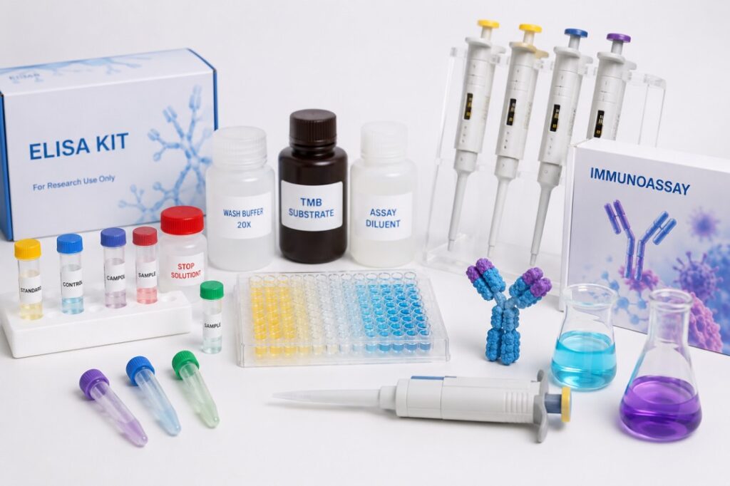
Introduction
The Immunology category supports the detection, measurement, and analysis of immune-related proteins, antibodies, cytokines, receptors, and cell populations. ELISA, Western blotting, and flow cytometry use different analytical principles, but each method depends on carefully selected antibodies, controls, buffers, blocking materials, labels, and detection reagents.
These techniques help researchers investigate immune signalling, inflammation, infectious diseases, autoimmune conditions, cancer biology, vaccine responses, and therapeutic targets. Selecting compatible products for each stage improves sensitivity, reduces non-specific signals, and makes experimental results easier to reproduce.
How Does ELISA Support Immunology Research?
Enzyme-linked immunosorbent assays are widely used to detect or quantify soluble biological targets in serum, plasma, cell-culture supernatants, tissue extracts, and other samples.
Depending on the format, an ELISA may measure:
- Cytokines and chemokines
- Antibodies and antigens
- Growth factors
- Hormones
- Inflammatory biomarkers
- Pathogen-associated proteins
- Therapeutic antibodies
- Cell-signalling molecules
The assay normally relies on specific binding between an antibody and its target. This interaction is converted into a measurable colour, fluorescent, or luminescent signal.
Researchers must consider the expected target concentration, sample type, required detection range, available plate reader, and number of samples before selecting an assay.
Which Products Influence ELISA Performance?
Successful ELISA workflows require more than a capture and detection antibody. Plates, standards, controls, buffers, enzyme conjugates, substrates, and wash solutions all affect the final result.
Products within Western ELISA can support antibody-based protein detection and related assay preparation. Researchers should match every reagent to the assay format and confirm compatibility with the chosen enzyme or label.
Important selection factors include:
- Antibody specificity
- Standard concentration range
- Sample dilution requirements
- Blocking-buffer composition
- Washing efficiency
- Incubation temperature
- Substrate sensitivity
- Signal-development time
Poor washing may leave unbound antibodies in the well and increase background. Excessive washing or unsuitable buffers may weaken the required immune complexes.
Why Is Blocking Important in ELISA?
After a plate surface has been coated, unoccupied binding sites must usually be covered to reduce unwanted attachment of antibodies or sample proteins.
Suitable Blocking Reagents help reduce non-specific binding and improve the difference between positive and negative signals. The blocking material should not interfere with the target, antibodies, enzyme conjugate, or detection chemistry.
A blocker that performs effectively for one assay may be unsuitable for another. Milk-based blockers, protein solutions, and synthetic formulations can interact differently with antibodies and phosphorylated targets. Small optimisation studies can identify the concentration and incubation time that produce the clearest signal.
How Does Western Blotting Identify Proteins?
Western blotting separates proteins according to size before transferring them from a gel to a membrane. Antibodies are then used to detect a selected target.
This technique can help researchers investigate:
- Protein expression
- Molecular weight
- Protein processing
- Isoforms
- Signalling changes
- Antibody specificity
- Treatment responses
- Relative protein abundance
Products within Blotting Units, Membranes, Papers and Reagents support protein transfer and membrane-based detection. The membrane type, transfer method, buffer composition, current, voltage, and transfer duration must match the protein and gel system.
Incomplete transfer may produce a weak signal, while excessive transfer can allow smaller proteins to pass through the membrane.
Why Are Protein Markers Needed?
Molecular-weight markers help researchers estimate the size of detected protein bands and monitor electrophoretic separation.
Protein Markers for Western Blotting should cover the expected molecular-weight range of the target. A marker that does not include bands near the target size provides limited support for interpretation.
Pre-stained markers can also help confirm gel progression and transfer orientation. However, researchers should check whether the marker is compatible with the selected membrane, detection chemistry, and imaging system.
How Can Western Blot Background Be Reduced?
High background may result from excessive antibody concentration, insufficient blocking, incomplete washing, contaminated buffers, or overexposure during detection.
Researchers can improve signal clarity by:
- Optimising primary and secondary antibody dilutions
- Increasing washing consistency
- Selecting a compatible blocking reagent
- Preparing fresh buffers
- Preventing membrane drying
- Controlling exposure time
- Using clean incubation containers
- Including suitable negative controls
Weak bands may require increased sample loading or improved antibody conditions, but simply increasing antibody concentration can also raise background noise.
How Does Flow Cytometry Analyse Immune Cells?
Flow cytometry measures physical and fluorescent properties of individual cells as they pass through a focused detection system. It can analyse thousands of cells within a short period and distinguish multiple immune-cell populations in one sample.
Flow cytometry can support:
- Immune-cell identification
- T-cell and B-cell analysis
- Cell viability measurement
- Proliferation studies
- Cytokine detection
- Receptor-expression analysis
- Apoptosis research
- Cell-cycle studies
Products within Kits for Flow Cytometry (FC) can support labelled cell analysis and proliferation-related measurements. The chosen kit should match the sample type, instrument lasers, available filters, cell number, and intended biological endpoint.
Why Does Fluorophore Selection Matter?
Each fluorescent label has specific excitation and emission characteristics. A fluorophore must be compatible with the instrument’s laser and detection channels.
In multicolour experiments, researchers should choose labels with limited spectral overlap. Compensation controls are required when fluorescence from one detector contributes to another channel.
Bright fluorophores are often assigned to weakly expressed targets, while dimmer labels may be suitable for abundant markers. Researchers must also consider photostability, fixation compatibility, and whether the fluorophore remains stable during the full procedure.
How Do Immunodetection Substrates Generate Signals?
Antibody-based methods often depend on enzyme substrates that produce visible or measurable signals after interacting with labelled antibodies.
Products within Substrates for Immunodetection can support colourimetric, fluorescent, or chemiluminescent detection. The selected substrate must match the enzyme conjugate and available reader or imaging system.
Highly sensitive substrates can reveal low-abundance targets, but excessive development may produce saturation. Once a signal is saturated, differences between samples become difficult to measure accurately.
Which Controls Improve Reliability?
Controls help determine whether a signal represents genuine target detection or technical interference.
Useful controls include:
- Unstained cells
- Single-colour controls
- Isotype controls
- Secondary-only controls
- Positive and negative samples
- Loading controls
- Blank ELISA wells
- Standard curves
- Viability controls
Researchers should apply the same sample preparation, incubation, washing, and analysis settings to comparable samples. Consistent documentation also makes unexpected results easier to investigate.
Conclusion
Immunology products support ELISA, Western blotting, and flow cytometry by enabling specific recognition and measurement of proteins, antibodies, biomarkers, and immune-cell populations.
ELISA provides efficient analysis of soluble targets, Western blotting combines protein separation with antibody detection, and flow cytometry examines individual cells using fluorescent markers. Although these methods differ, all require compatible antibodies, blocking materials, controls, substrates, and carefully managed experimental conditions.
Selecting products according to the target, sample type, detection system, and instrument improves signal quality, reduces background interference, and supports more dependable immunology research.